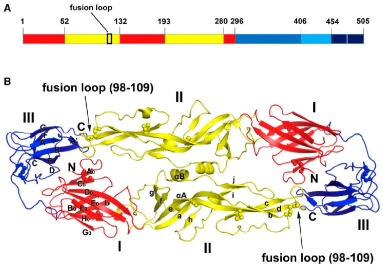
Figure 2.
Structure of the E protein of ZIKV. (A) Domain organization of ZIKV E protein. Domain I, II and III are schematically indicated with red, yellow, and blue bars, respectively. (B) Dimer structure of the E protein of ZIKV. Domain I, II and III follow the same color scheme as Panel A and the position of fusion loop is indicated with an arrow. The locations of epitopes for various antibodies are indicated with spheres. (From [54] with permission from Elsevier).

