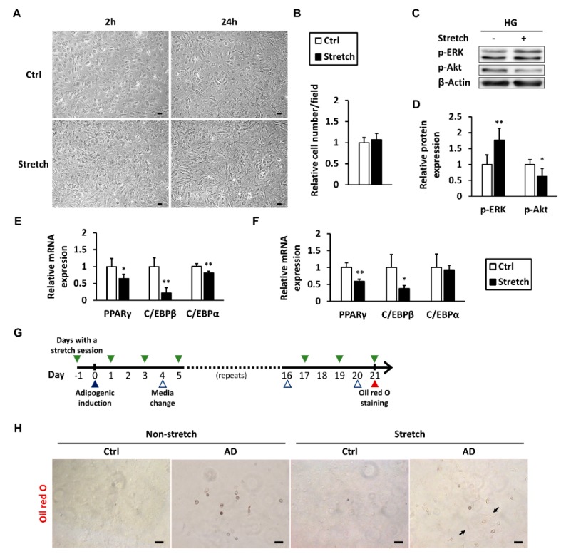
Figure 5.
Mechanical stretch modulates ERK and Akt signaling and represses adipogenic differentiation. Tenocytes in high-glucose conditions were subjected to a 2-h mechanical stretch session. (A) Cell morphology was observed using a light microscope 2 and 24 h after the session began, scale bar = 50 μm; (B) Cell number was counted in three random visual fields from each of the three culture wells at the 24-h time point; 6 h after the stretch began, p-ERK and p-Akt in tenocytes were (C) immunoblotted and (D) quantified (n = 4); The mRNA expression of adipogenic markers was analyzed at (E) 2- and (F) 6-h time points; Data are presented as the mean ± SD (n = 4). Statistical significance is shown as * p < 0.05 or ** p < 0.01 compared to the non-stretched control group; (G) Timeline of the adipogenic differentiation protocol for tenocytes with regular exposure to mechanical stretch; (H) Adipogenesis was examined using Oil Red O staining on the 21st day of induction. Arrows indicate the stretched tenocytes retained their spindle shape. AD: adipogenic differentiation medium. Ctrl: culture medium. Scale bar = 50 μm.

