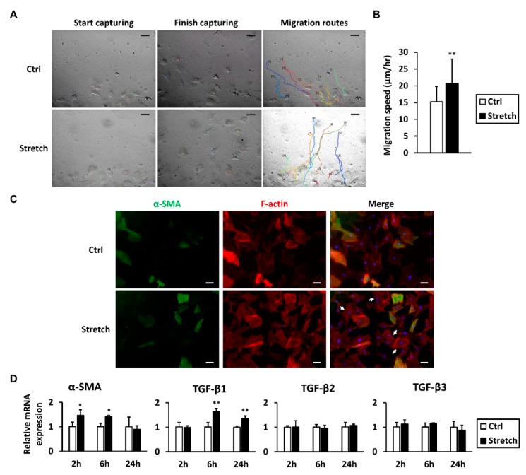
Figure 6.
Mechanical stretch promotes tenocyte characteristics. (A) Motility of tenocytes subjected to mechanical stretch was analyzed using a wound healing assay. Wounds were created before stretch, and wound closure was monitored in real time up to 24 h. Migration routes were delineated using time-lapse videos. Each line represents the path of a different cell; (B) Migration speed of randomly chosen cells was calculated (n = 70); (C) F-actin and α-SMA were fluorescently labeled immediately after the stretch session. After stretch, F-actin in α-SMA-negative tenocytes were sharper than those in the non-stretched control group (arrows); (D) The mRNA expression levels of α-SMA and TGF-βs were measured 2, 6, and 24 h after stretch began (n = 4). Data are presented as the mean ± SD. Statistical significance is shown as * p < 0.05 or ** p < 0.01 compared to the non-stretched control group. Scale bar = 50 μm.

