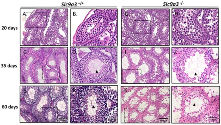
Figure 5.
The testicular sections of 20-, 35-, and 60-day-old Slc9a3 knockout mice. Comparison of the testicular sections of WT (A–F) and Slc9a3−/− (G–L) mice according to H&E staining; (B,D,F,H,J,L) enlarged images from the areas boxed by a black dashed box in (A,C,E,G,I,K); Scale bar = 100 μm (E,K) and 50 μm (F,L); Elongated spermatids (arrowhead) observed in the ducts of the seminiferous tubules (D,F,J) but absent in (L).

