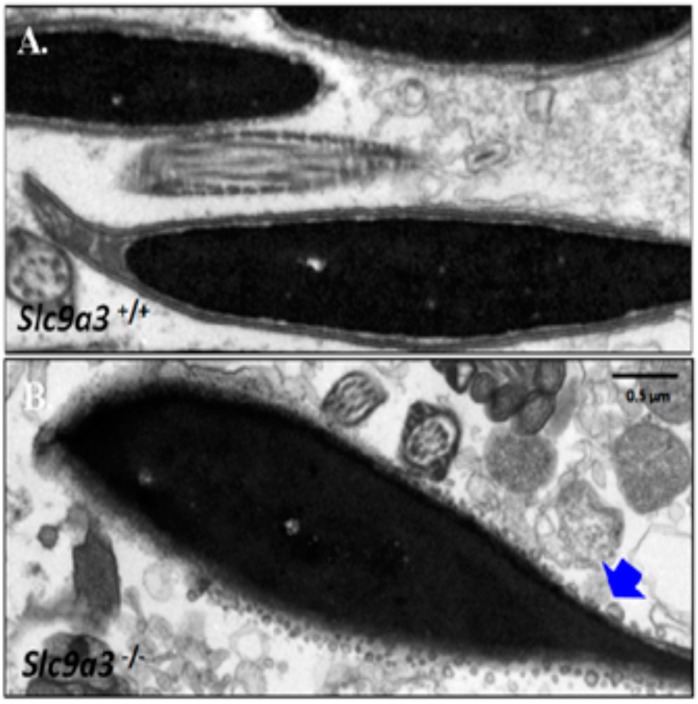
Figure 7.
Ultrastructural defects of the sperm acrosome from Slc9a3–/– mice. Sperm isolated epididymis of Slc9a3+/+ (A) and Slc9a3−/− (B) mice. Electron micrograph showing the ultrastructure of sperm of 35-day-old WT (upper panel) and Slc9a3−/− male mice (lower panel). Arrow indicates the acrosome defects in sperm from Slc9a3−/− male mice. Scale bar = 0.5 μm.

