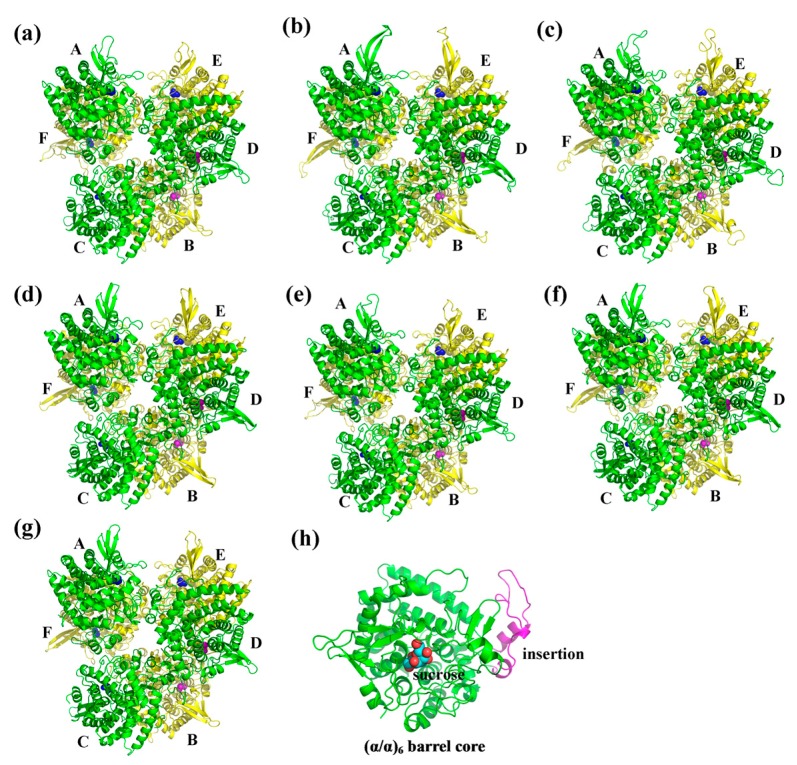
Figure 6.
Cartoon representation of the predicted three-dimensional structure models of CaNINV1–6. (a) CaNINV1; (b) CaNINV2; (c) CaNINV3; (d) CaNINV4; (e) CaNINV5; (f) CaNINV6; and, (g) NINV protein from Anabaena; (h) the monomer of CaNINV1. The six subunits are sequentially labeled as A–F. The spherical structures indicate sucrose molecules. The image was generated using the PyMOL program (Schrödinger, Inc., New York, NY, USA).

