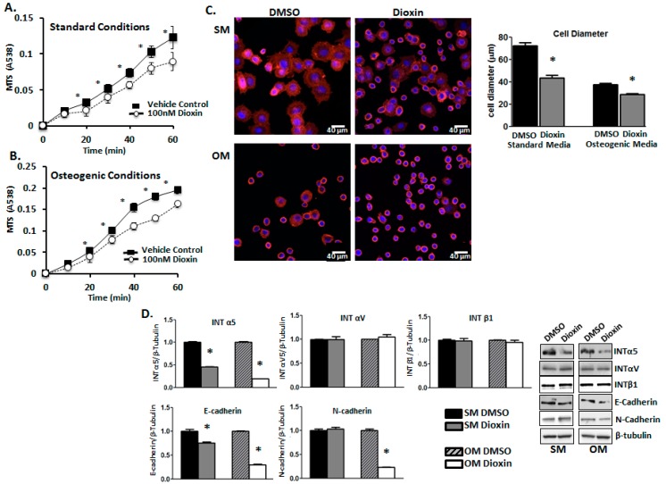
Figure 3.
Dioxin reduces cell adhesion in both un-induced and differentiating MG-63 cells. Cell adhesion rates were quantified after a dioxin pre-treatment period of 3 days under either standard (A) or osteogenic conditions (B). Significance is shown relative to vehicle control-treated cells under both standard and osteogenic conditions; (C) Visualization of cell morphology. Dioxin exposure significantly decreased the proportion of flattened cells under both standard and osteogenic conditions, whereas the proportion of rounded cells was increased in response to dioxin treatment. Rhodamine-bound F-actin is shown in red, whereas nuclei are shown in blue; (D) Integrin (INT) α5 and E-cadherin protein expression levels were significantly decreased in dioxin-exposed cells under both standard and osteogenic conditions, whereas INTαV and INTβ1 were unchanged. N-cadherin levels were decreased only in differentiating dioxin-treated cells. * p < 0.05 relative to 0 nM dioxin under standard or osteogenic conditions. Error bar means ± SEM.

