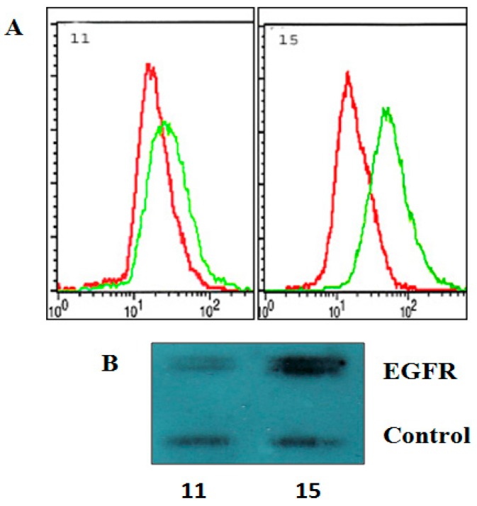
Figure 2.
Membrane expression of EGFR on 11 HGG and 15 HGG cells. For flow cytometry determination (A), cells were stained with a PE-conjugated anti- EGFR, or a PE-labelled isotype Mouse IgG2B-κ control antibody (red line) and of EGFR was (green line) analyzed as described in Materials and methods; For Western blot analysis (B), cell lines were lysed, electrophoresed, and immunoblotted with a EGFR antibody. Membranes were reprobed with an actin antibody as a loading control.

