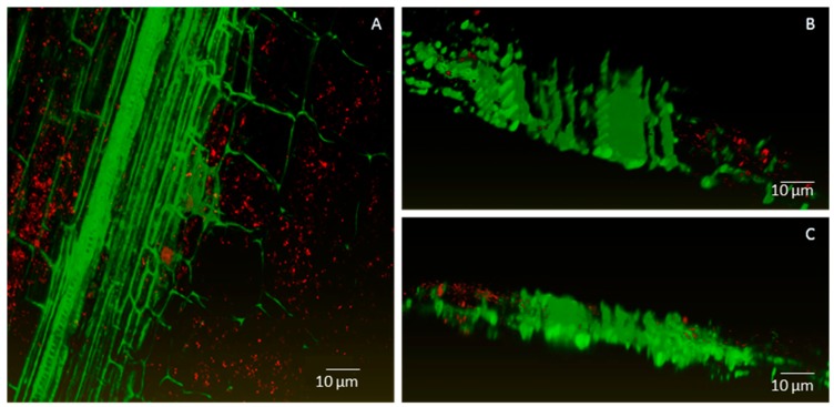
Figure 4.
Confocal images of combined m-Cherry fluorescence (red) and plant autofluorescence (green) showing root colonisation by Methylobacterium strain Cp3. Maximum intensity projection (A) and volume rendering (B,C) where Cp3-mCherry is localized intracellularly in root cortex of Crotalaria pumila. Confocal stack thickness is 58 μm and was acquired with the Ultra VIEW VoX (PerkinElmer, Zaventem, Belgium) using the CFI Plan Apochromat VC objective 20.0 × 0.75. Z-step was 1 μm. Three-dimensional models were created with the software Amira 6.0.1 (FEI software, Hillsboro, OR, USA).

