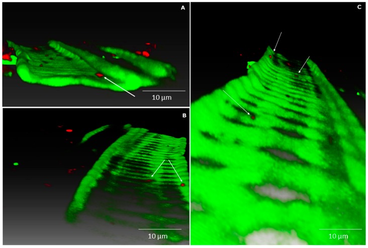
Figure 5.
Confocal images with combined mCherry fluorescence (red) and plant autofluorescence (green). Volume rendering (A–C) of Methylobacterium sp. Cp3-mCherry colonising the xylem vessels in the stem of Crotalaria pumila growing in medium supplemented with Zn and Cd. White arrows indicate strain Cp3. Confocal stack has a thickness of 54 μm, acquired with a Ultra VIEW VoX (PerkinElmer) using the CFI Plan Fluor objective 40.0 × 0.75. Z-step was 1 μm. Three-dimensional models were created with the software Amira 6.0.1 (FEI software, Hillsboro, OR, USA).

