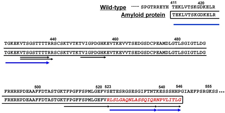
Figure 2.
Putative primary structure of amyloid fibril protein in a Japanese Aα-chain amyloidosis patient [14]. Amyloid protein in this patient is assumed to be composed of both wild-type sequence (residues 411–522) and an additional fragment (red italic letters) induced by the frameshift variant (residues 523–546). Black arrows denote tryptic peptides detected at laser microdissection (LMD) isolation of glomerular amyloid, and blue arrows denote Arg-C peptides detected at in-gel digestion on SDS-PAGE analysis of duodenal amyloid.

