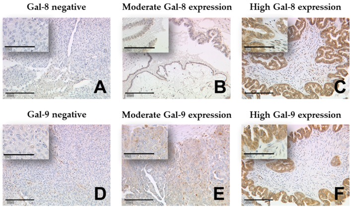
Figure 1.
Detection of galectin-8 and -9 using immunohistochemistry (IHC). Gal-8 staining (A–C) and Gal-9 staining (D–F) was predominantly present in the cytoplasm of ovarian cancer cells but not in the peritumoral stroma. Representative photomicrographs are shown. There is a 10× magnification for the outer pictures and 25× for the inserts. Scale bar in (A) equals 200 μm (outer pictures) and 100 μm (inserts). Gal: galectin.

