
Fig. 2.
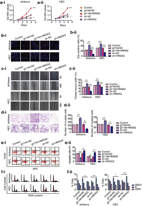
HMGA2 plays an oncogenic role in Ishikawa and HEC-1A cell lines. a and b CCK-8 and EdU assays were used to detect the effect of HMGA2 expression on the proliferation of Ishikawa and HEC-1A cells. c Effects of different levels of HMGA2 on the migration of Ishikawa and HEC-1A cells were evaluated using a wound healing assay. d Transwell assays were used to detect cellular invasion. Representative images and accompanying statistical plots are presented. e The effects of different levels of HMGA2 on apoptosis were verified via flow cytometry. f Effect of HMGA2 on cell cycle distribution in Ishikawa and HEC-1A cell lines. Data are presented as the mean ± SEM (n = 3 per group), *P < 0.05, **P < 0.01, *** P < 0.0001 vs. the NC group