
Fig. 6.
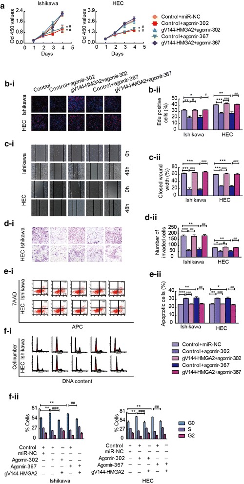
miR-302a-5p/367-3p suppresses the malignant behaviour of endometrial carcinoma cells by targeting HMGA2. a and b CCK-8 and EdU assays were used to detect proliferation in Ishikawa and HEC-1A cell lines after co-transfection with miR-302a-5p/367-3p and HMGA2-expression constructs. c A wound healing assay was performed to investigate the migratory ability of the cells. d Quantification of cell invasion in the groups with different expression levels of miR-302a-5p/367-3p and HMGA2. Representative images and statistical plots are presented. e Flow cytometric analysis of apoptosis in Ishikawa and HEC-1A cells expressing various levels of miR-302a-5p/367-3p and HMGA2. f Representative flow diagrams and graphs showing cell cycle distribution in the groups with different levels of miR-302a-5p/367-3p and HMGA2. Data are presented as the mean ± SEM, (n = 3). *P < 0.05, ** P < 0.01 vs. control vector + miR-NC, #P < 0.05, ## P < 0.01, ### P < 0.0001 vs. control vector + agomir-302a-5p/367-3p