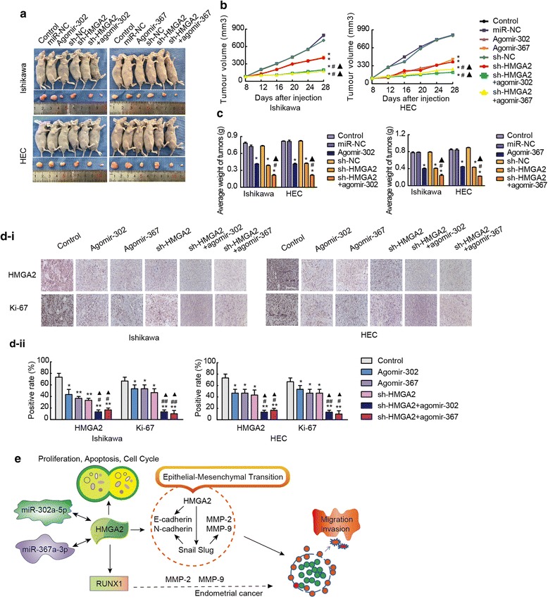
Fig. 7.

In vivo tumour xenografts. a Nude mice bearing tumours composed of cells from the respective groups; sample tumours from each group are shown. b and c Tumour volume was measured every 4 days after injection. Twenty-eight days later, the mice were sacrificed, and the tumours were excised and weighed. *P < 0.001 vs. the control group, #P < 0.001 vs. the agomir group, ▲P < 0.001 vs. the sh-HMGA2 group. d Expression levels HMGA2, Ki-67. Positive cells are stained brown. Quantified protein levels (area %) in representative xenograft tumours from each treatment group are shown. Data are presented as the mean ± SEM. (n = 3 per group). *P < 0.05, ** P < 0.01, ***P < 0.0001 vs. the control group, #P < 0.05, ## P < 0.01, ### P < 0.0001 vs. the agomir group, ▲P < 0.05, ▲▲ P < 0.01, ▲▲▲P < 0.0001 vs. the sh-HMGA2 group. e A working model showing the interaction between miRNAs and HMGA2 in Ishikawa and HEC-1A cells
