
Fig. 3.
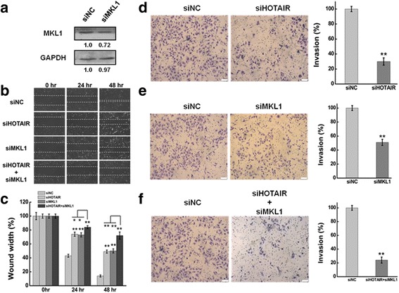
a Western blots analysis of the MKL1 protein expression at 48 h after transfected with siMKL1 (siMKL1-I, siMKL1-II and siMKL1-III) or siNC. GAPDH was used as an internal control. b The effect of knockdown HOTAIR or/and MKL1 on cell migration was determined by wound healing assay. c Quantification of the wound healing assay. d, e, f The effect of HOTAIR or/and MKL1 inhibition on cell invasion was determined in a Boyden chamber assay. And the number of cells on the underside of the filter was determined and significantly (P < 0.05) changed invasion is indicated. Data are presented as means ± S.D. and represent results from three independent experiments. Statistically significant differences are indicated: *, P < 0.05; **, P < 0.01 (Student’s t test)