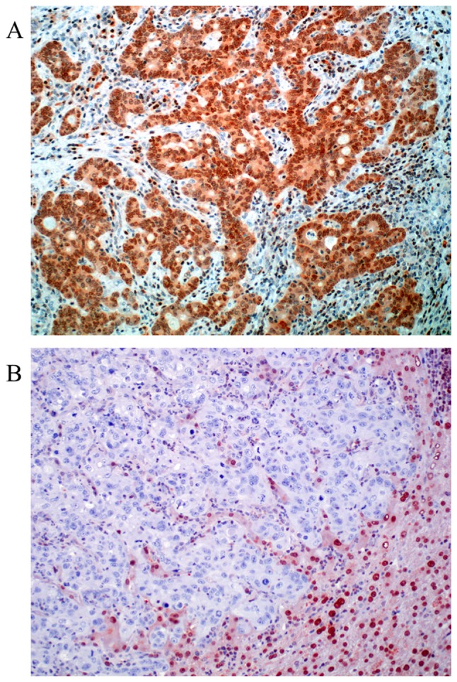
Figure 2.

Representative examples of O6-methylguanine DNA methyltransferase protein expression in colorectal liver metastasis. (A) Score 0, negative staining-<10% of the nuclei are stained. (B) Score 1, >10% percent of the nuclei are positive. Note that in negative cases, stromal cells, lymphocytes and hepatocytes are still nuclear positive. Magnification, ×200.
