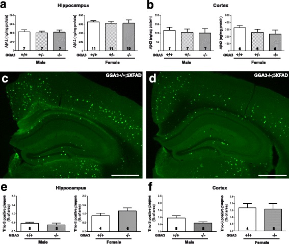
Fig. 3.

GGA3 deletion does not increase levels of Aβ42 and amyloid plaques in young 5XFAD mice. a-b Human Aβ42 levels were measured by ELISA (ng/mg of total protein) (Invitrogen) in hippocampus (a) and cortex (b) GuHCl extracts from of 4 months old GGA3WT;5XFAD, GGA3Het;5XFAD and GGA3KO;5XFAD male and female mice. Levels of Aβ42 were similar in all genotypes at 4 months of age. Total number of mice analyzed is indicated within bars. One-way ANOVA with Fisher’s LSD post hoc tests was applied to each genotype group. c-d Coronal brain sections from 4 months old GGA3WT;5XFAD and GGA3KO;5XFAD male and female mice were used to analyze amyloid plaques by Thioflavin-S staining. Representative images show amyloid plaques from 4 months old female GGA3WT;5XFAD (c) and GGA3KO;5XFAD (d) mice. Scale bar is 500 μm. e-f The graphs represent the percentage area occupied by amyloid plaques stained with Thioflavin-S in hippocampus (e) and cortex (f) of GGA3WT;5XFAD and GGA3KO;5XFAD male and female mice at the age of 4 months. GGA3 deletion does not affect amyloid burden in 4 months old 5XFAD mice. Total number of mice in each group is indicated within bars. All graphs represent mean ± SEM. Unpaired t-test with Welch’s correction was performed
