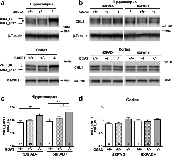
Fig. 4.

BACE1-mediated cleavage of CHL1 is increased in the hippocampus of young GGA3KO and 5XFAD;GGA3KO mice. a Representative immunoblots of hippocampus (top) and cortex (bottom) homogenates from BACE1+/+, BACE1+/−, and BACE1−/− mice using anti-N-terminal CHL1 antibody (AF2147). Brain homogenates with reduced BACE1 exhibit increased intensity of full-length CHL1 (CHL1_FL) at ~ 185 kDa and a reduction (BACE1+/−) or absence (BACE1−/−) of BACE1-cleaved N-terminal CHL1 (CHL1_βNTF) at ~ 165 kDa. An ~ 175 kDa band between CHL1_FL and CHL1_βNTF may present an alternative splicing isoform or glycosylated CHL1 in hippocampus. b Representative immunoblots of CHL1_FL and CHL1_βNTF levels in hippocampus (top) and cortex (bottom) homogenates from 4 months old GGA3WT, GGA3Het, GGA3KO, GGA3WT;5XFAD, GGA3Het;5XFAD and GGA3KO;5XFAD mice. c-d Graphs represent the ratio of CHL1_βNTF to CHL1_FL in hippocampus (c) and cortex (d) homogenates in the six different genotypes. GGA3 deletion significantly increased CHL1_βNTF/CHL1_FL ratio in the hippocampus of non-5XFAD and 5XFAD mice, whereas no changes were observed in the cortex. All graphs represent mean ± SEM. One-way ANOVA with Fisher’s LSD post hoc tests was applied to each genotype group. * p < 0.05, ** p < 0.01
