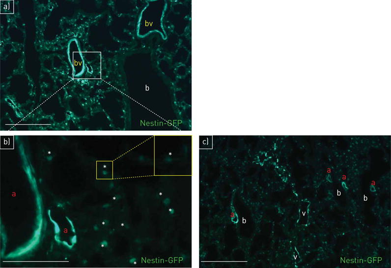
FIGURE 1.

Localisation of nestin-GFP (green fluorescent protein) in lung vasculature. a) Nestin-GFP+ cells are detectable in pulmonary blood vessels (bv); bronchioli (b) are nestin-GFP−; day 1. b) (detail of panel a at higher magnification) Nestin-GFP+ cells in arteries (a) and capillaries (asterisks mark the lumen); day 1. c) Nestin-GFP+ cells in arteries (a) and veins (v); bronchioli (b) are nestin-GFP−; day 1. Scale bars: a, c) 100 μm; b) 50 μm.
