FIGURE 2.

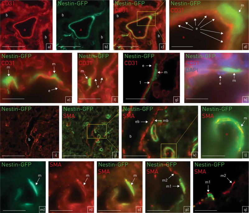
Nestin-GFP (green fluorescent protein) is co-localised with the smooth muscle cell marker smooth muscle actin (SMA), but not with the endothelial cell marker CD31. a–h) Nestin-GFP (green) and CD31 (red) (a–d, h: day 1; e–g: adult). a–d) Artery (a): nestin-GFP is not co-localised with CD31. Bronchioli (b) are nestin-GFP− (a: CD31; b: nestin-GFP; c: merge). d) (detail of panel c at higher magnification) The nestin-GFP+ vascular smooth muscle cell (VSMC) (m) is outside of CD31+ endothelial cells (e). e, f) Single nestin-GFP+ VSMCs (m). They are not co-localised with CD31+ endothelial cells (e). g) (confocal microscopy) Clear separation (labelled by “1”) between the CD31+ layer of endothelial cells (e) and the nestin-GFP+ layer of VSMCs (m). h) Additional staining with 4′,6-diamidino-2-phenyl-indole (DAPI); DAPI clearly reveals the nuclei from CD31+ endothelial cells (e). m: nestin-GFP+ VSMC. i–q) Nestin-GFP (green) and SMA (red) (i–o: day 1; p, q: adult). i, j) Nestin-GFP+/SMA+ arteries (a) are adjacent to a nestin-GFP−/SMA+ bronchiolus (b) (i: SMA; j: SMA and nestin-GFP). k) (detail of panel j at higher magnification) Nestin-GFP+/SMA+ VSMCs of arteries (a); smooth muscle cells of bronchioli (mb) are SMA+/nestin-GFP−. The epithelium of bronchioli (eb) is SMA−. l) (detail of an artery of panel k at higher magnification) Nestin-GFP+/SMA+ VSMCs (m) of an artery (a). m–o) (m: nestin-GFP; n: SMA; o: merge) Nestin-GFP+/SMA+ VSMC (m). SMA is present in the cytoplasm (n: red), Nestin-GFP is located to the cytoplasm (o: yellow) and the nucleus (o: green). p, q) Only a subpopulation of VSMCs is nestin-GFP+. Nestin-GFP+/SMA+ VSMCs (m1); nestin-GFP−/SMA+ VSMCs (m2) (q: confocal microscopy). Scale bars: a–c) 75 μm; d, m, n, o) 15 μm; e–g, q) 20 μm; h, l, p) 25 μm; i, j) 100 μm; k) 50 μm.
