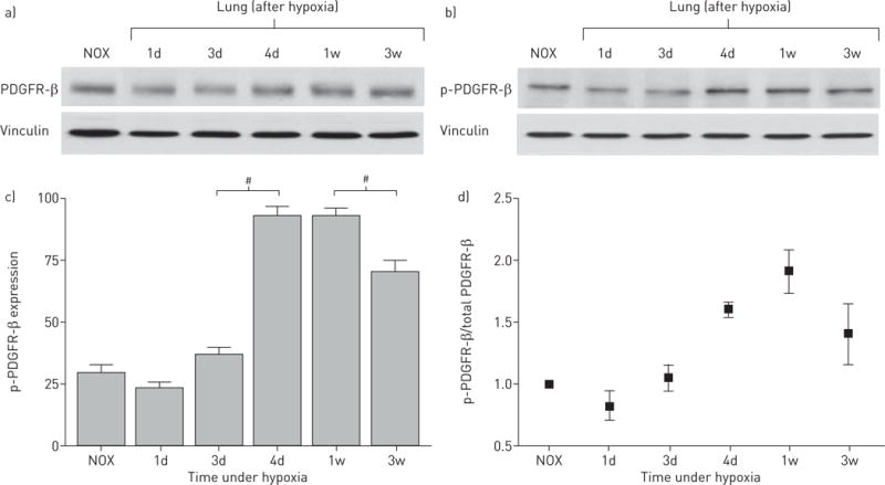
FIGURE 4.

Platelet-derived growth factor receptor β (PDGFR-β) expression in hypoxic mouse lung samples. a) PDGFR-β Western blot shows only a slight increase in PDGFR-β expression during hypoxic exposure towards 3 weeks (30 μg membrane protein). b) Phospho (p)-PDGFR-β Western blot shows a peak of activated (phosphorylated) PDGFR-β after 4 days hypoxia (30 μg membrane protein). c) Densitometric quantification of p-PDGFR-β expression. Bars show mean±SEM scores from three assessments. #: p<0.01 indicates significant differences in p-PDGFR-β expression between 3 and 4 days as well as between 1 and 3 weeks. Vinculin was used as loading control. d) PDGFR-β activity expressed as p-PDGFR-β/PDGFR-β ratio. NOX: normoxia; 1d: 1 day; 3d: 3 days; 4d: 4 days; 1w: 1 week; 3w: 3 weeks.
