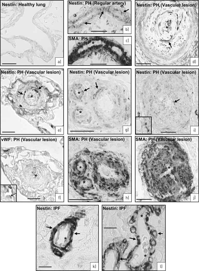
FIGURE 7.

Human lungs. a) Healthy lung. No nestin-expressing cells. b–j) Pulmonary hypertension (PH) lungs. b, c) Serial sections of a regular artery stained for nestin (b: arrows) and smooth muscle actin (c: asterisks mark SMA− endothelial cells). d) Artery with complex lesion stained for nestin (arrows). e–j) Serial sections of three different arteries with complex lesions stained for nestin (e: arrows) and von Willebrand factor (vWF) (f, inset: arrows) as well as nestin (g, i: arrows) and SMA (h, j: asterisks mark SMA negative-endothelial cells). k–l) Idiopathic pulmonary fibrosis (IPF) lung. Nestin immunoreactivity in smooth muscle cells (arrows) of the arterial media. Asterisks mark lack of nestin staining in endothelial cells. Scale bars: a–d, g–j) 50 μm; e, f, k, l) 10 μm.
