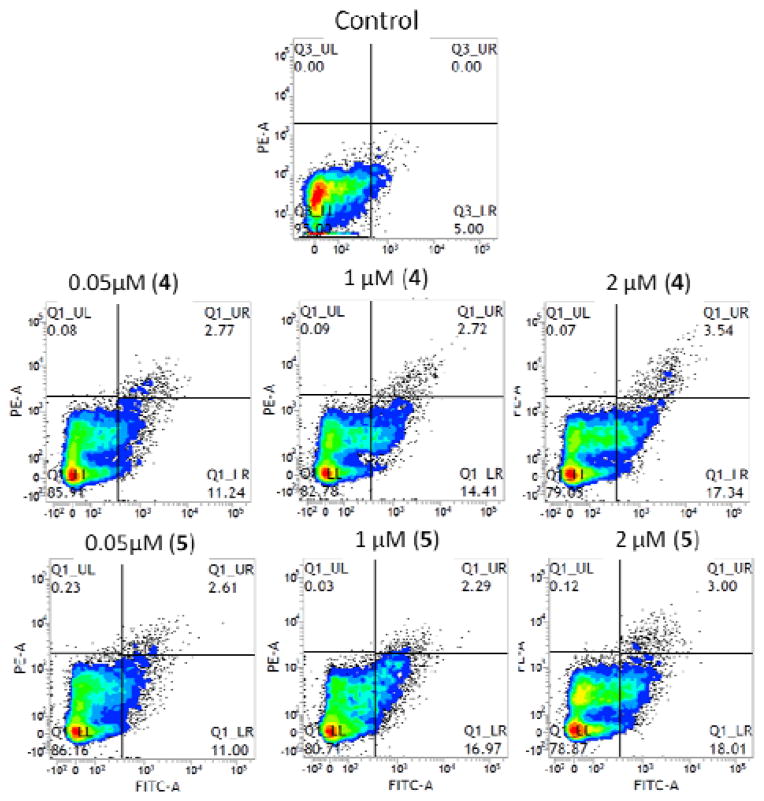
Figure 9.
Detection of apoptosis in human T98G brain tumor cells. Cells treated with complexes 4, and 5 at various concentrations (0.05–2 μM) were suspended in binding buffer. Annexin V-FITC and propidium iodide solution were added and cell solutions were incubated for 15 min at room temperature in the dark. Flow cytometric analysis was performed within 1 h. Each number represents the percentage of cells in each quadrant.

