Fig. 6.

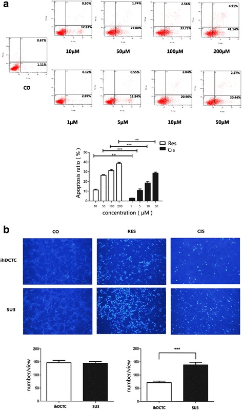
Res have stronger ablity to induce apoptosis of ihDCTC cells then Cis. a Flow cytometry was used to detect different concentration gradient’s Res(10、50、100、200 μM)and Cis(1、5、10、50 μM) deal with ihDCTC for 48 h.Left lower quadrant was normal cells, upper right quadrant was late apoptotic cells, early apoptosis cells at the lower right quadrant. b 100 μM Res and 10 μM Cis were treated with ihDCTC and SU3 48 h and Hoechest33342 for 30 min respectively. Apoptotic cell nucleus which was stained blue were obversed under the purple excitation light by Fluorescence microscope. Compared with control groups. **P < 0.01
