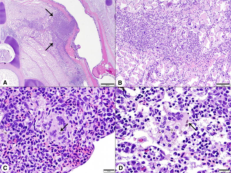
Figure 2.

Histologic findings in white sturgeon fingerlings challenged by intramuscular injection with Veronaea botryosa at 13 °C. A Low magnification photomicrograph of an irregular area of granulomatous inflammation (arrows) radiating into skeletal muscle at the injection site. H&E stain, Bar = 500 μm. B Higher magnification image of muscle lesion. Inflammatory infiltrates were dominated by macrophages, with smaller numbers of multinucleated giant cells and lymphocytes. H&E stain, Bar = 100 μm. C Spleen, multinucleated giant cell containing pigmented hyphal fragments (arrow). H&E stain, Bar = 20 μm. D Kidney, giant cells with hyphal fragments (arrow) surrounded by hematopoietic tissue and renal sinusoids. H&E stain, Bar = 20 μm.
