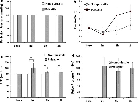
Fig. 2.

Graphs showing the mean perfusion pressure, flow and EEP of 30 s data at each specified time period. Initially, the mean perfusion pressures in both groups showed no difference (a). The mean perfusion flow of the SDS solution increased more rapidly in the pulsatile group (b). The baseline EEP (Energy Equivalent Pressure), before pulsatile flow was applied, showed no difference between the two groups. After pulsatile flow was applied, the EEP was dramatically increased (c). Pulse pressure shown for comparison with EEP (d). *p < 0.05
