Fig. 4.

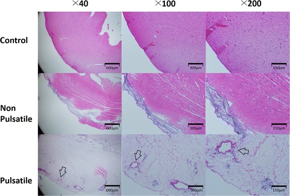
Hematoxylin and Eosin stain of the rat hearts after 2 h of decellularization showed more profound decellularization in the pulsatile group. The vascular structures are visible in the pulsatile group (arrow)

Hematoxylin and Eosin stain of the rat hearts after 2 h of decellularization showed more profound decellularization in the pulsatile group. The vascular structures are visible in the pulsatile group (arrow)