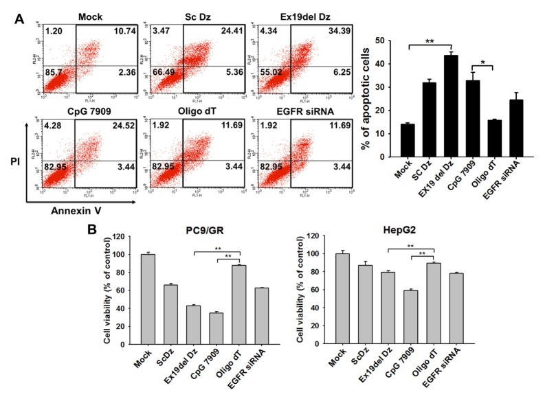
Fig. 3.
DNA containing the CpG site causes apoptotic cell death in lung cancer cells. (A) 100 nM individual oligo DNA was transfected into lung cancer cells (PC9/GR). After 48 h of incubation, cell fate was analyzed by flow cytometry (FACS analysis). The graph represents the estimated percentage of apoptotic cells determined in the FACS analysis. *P < 0.05 and **P < 0.01 (B) Lung cancer cells (PC9/GR) and liver cancer cells (HepG2) were seeded onto 24-well plates, and each oligo DNA (100 nM final concentration) and EGFR siRNA (50 nM) were transfected into the cells followed by incubation for 48 h. Cell viability was assessed using the methylene blue assay. Data are the mean ± S.D. of three independent experiments; *P < 0.05 and **P < 0.01 vs. the oligo dT group.

