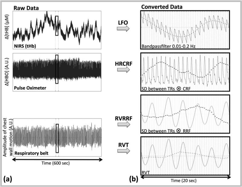
Figure 1. Steps for temporal and spatial analysis.
(a) The raw signals from the NIRS, Pulse Oximeter and Respiratory belt acquisition are shown in the first column. (b) Four methods were used to convert these raw signals into LFO noise regressors (dotted line in second column): 1) The NIRS LFO method which involved, direct measurements of LFOs in the periphery using NIRS (13), 2) the cardiac variation method (HRCRF) (5), 3) the respiratory variation method (RVRRF) (5) and 4) the respiration volume per time (RVT) method (4). The LFO noise regressors were then downsampled to the acquisition TR (0.4 s) and compared both temporally, and spatially (via their maximum correlation map).

