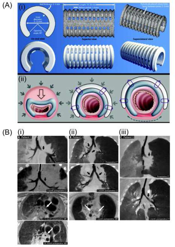
Figure 9.
(A) Computational image-based design of 3D-printed tracheobronchial splints. (B) Pre- and post-operative imaging of patients. (i) Preoperative (top) and 1-month postoperative (upper middle) CT images of patient 1. Postoperative MRI (lower middle) demonstrated presence of splint around left bronchus in patient 1 at 12 months and focal fragmentation of splint due to degradation at 38 months (bottom). (ii) Preoperative (top) and 1-month postoperative (upper middle) CT images of patient 2. Postoperative MRI (lower middle) demonstrated presence of splints around the left and right bronchi in patient 2 at 1 month. (iii) Preoperative (top) and 1-month postoperative (bottom) CT images of patient 3. Adapted with permission from [111].

