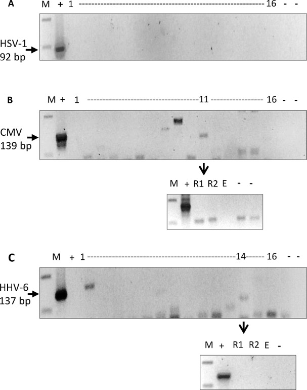
Fig 2. PCR amplification for 3 common herpesviruses.

PCR for HSV-1, CMV and HHV-6 was performed on randomly selected 16 white matter DNA that were EBV PCR positive. (A) None of the 16 DNA samples tested were positive for HSV-1 92bp fragment. (B) Similarly, none of these samples amplified CMV 139bp fragment. One sample (case 11) gave an amplification product of the expected size, but on repeat (R1 and R2), no amplification was seen. (C) All 16 DNA samples were also negative for HHV-6 137bp fragment. Sample 14 gave a weak band, but once again on repeating (R1, R2), it was found to be negative. [M: 100bp DNA marker; (-): negative control; (+): positive control DNA; E: empty well].
