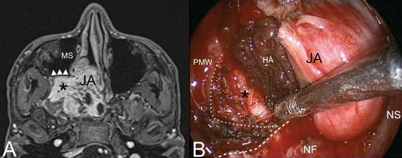
Fig. 2.

( A ) Axial contrast-enhanced T1-weighted magnetic resonance (MR). Juvenile angiofibroma (JA) with typical extension into the pterygopalatine and infratemporal fossa (black asterisk). The posterior wall of the maxillary sinus (MS) is pushed forward by the lesion (white arrowheads). ( B ) Identification of the dissection plane cutting the posterior periosteum of the posterior maxillary wall (PMW) (white dotted line). Inferior turbinate and medial maxillary wall have been removed to adequately expose the posterior maxillary wall. White dashed line: inferior limit of the medial maxillary wall. Abbreviations: HA, hemostatic agent; NF, nasal floor; NS, nasal septum.
