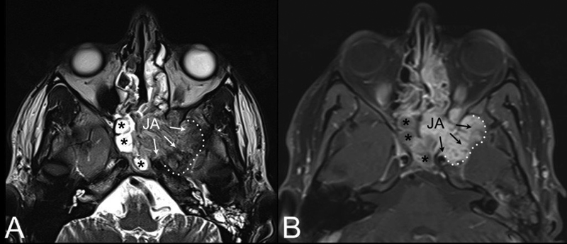
Fig. 4.

AxialT2-weighted ( A ) and contrast-enhanced T1-weighted magnetic resonance (MR) ( B ). Juvenile angiofibroma (JA) with intracranial extension (black arrows) into the middle cranial fossa through the pterygoid erosion and inferior orbital fissure. Extension to the anterior portion of the left cavernous sinus is also visible. Black asterisks: sphenoidal inflammatory tissue.
