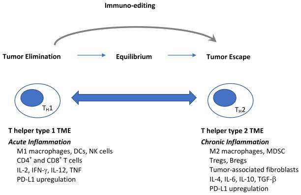
Figure 1. The immune system plays a role in breast tumor growth and progression, and also in breast tumor elimination.
Early in breast tumor development, the acute inflammatory response results in the production of interleukin-12 (IL-12) and interferon-γ (IFN-γ), establishing a T helper type 1 environment at the tumor site. During this phase, DCs mature, process tumor-associated antigens, and migrate to the tumor-draining lymph nodes to present antigen to naïve CD4+ and CD8+ T cells, resulting in an immune response that ultimately lyses tumor cells. This immune response initially results in complete tumor rejection. However, the pressure it imposes leads to the selection of tumor cell variants that escape the immune response. This process of immuno-editing establishes a state of equilibrium, or dormancy. As inflammation at the tumor site shifts from acute to chronic, the tumor microenvironment evolves to a T helper type 2 profile. A suppressive tumor microenvironment comprised of a complex community of tumor cells, immune cells and host stromal cells is established, and breast tumors grow and metastasize unchecked by the immune system. Abbreviations: TH1= T helper type 1; TH2=T helper type 2; TME= tumor microenvironment; DCs=dendritic cells, NK=natural killer, IL-2=interleukin-2; IFN-γ=interferon-γ; IL-12=interleukin-12; TNF=tumor necrosis factor; PD-L1=programmed death ligand-1; MDSC=myeloid-derived suppressor cells; IL-4=interleukin-4; IL-6=interleukin-6; IL-10=interleukin-10; TGF-β=transforming growth factor-β.

