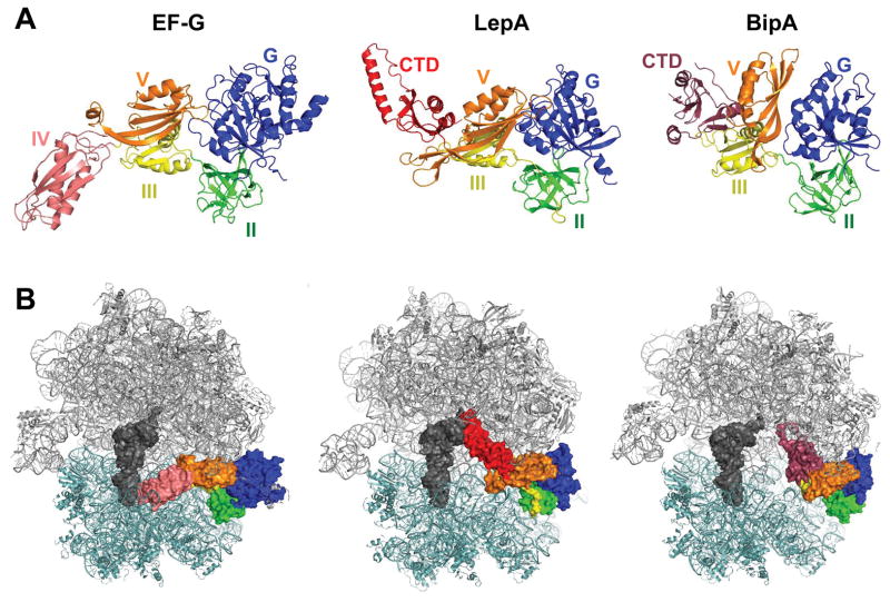
Figure 1. Homologous proteins EF-G, LepA, and BipA bind the ribosome similarly.
(A) Structures of EF-G, LepA, and BipA. Domains G (blue), II (green), III (yellow), and V (orange) are homologous among the factors. Unique domains include EF-G domain IV (pink), LepA C-terminal domain (red), and BipA C-terminal domain (raspberry). Images are based on PDB ID files 4V5F, 5J8B, and 4ZCI. (B) Structures of EF-G, LepA, and BipA bound to the 70S ribosome. 50S subunit, gray; 30S subunit, light teal; P-site tRNA; dark gray. Color coding of GTPase domains as in panel A. Images are based on PDB ID files 4V5F, 4W2E, and 5AA0.

