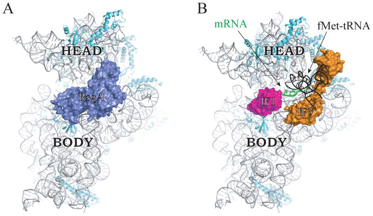
Figure 3. RsgA binds the 30S subunit in a way that occludes IF1, IF3, and initiator tRNA.
Interface view of the 30S subunit bound by RsgA (A) or by IF1, IF3, fMet-tRNA, and mRNA (B), as determined by cryo-EM studies (Hussain et al., 2016; Lopez-Alonso et al., 2017). 16S rRNA, gray; r proteins, cyan; RsgA, blue; IF1, magenta; IF3, orange; mRNA, green; fMet-tRNA, black. Head and body regions of the subunit are labeled. Images are based on PDB ID files 5NO3 and 5LMV.

