
Figure 3.
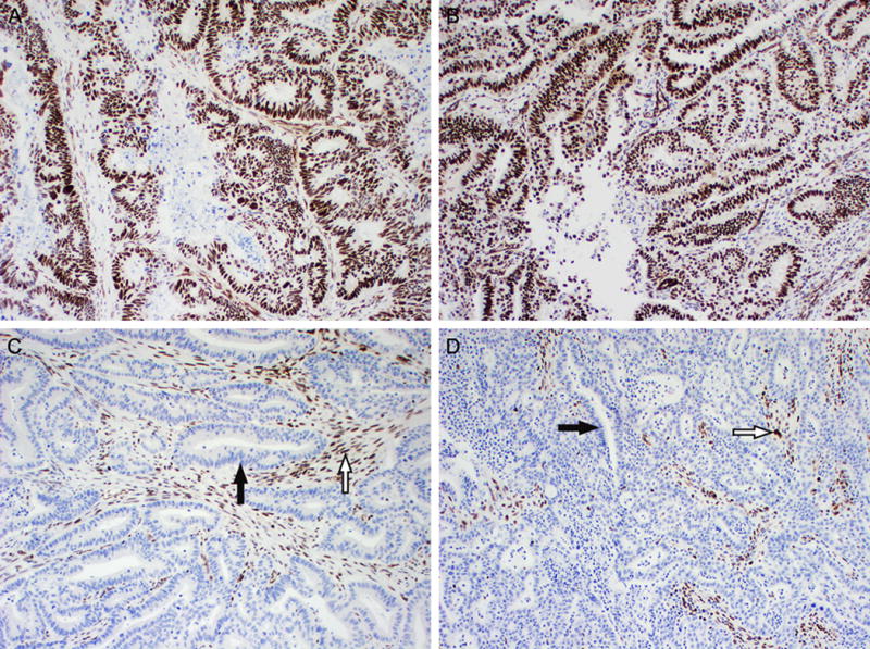
Examples of positive and negative MLH1 expression by immunohistochemistry. (A and B, 100×) Positive MLH1 expression. The nuclei of colonic adenocarcinoma (A) and endometrial endometrioid adenocarcinoma (B) exhibit diffuse strong staining. (C and D, 100×) Loss of MLH1 expression in colonic adenocarcinoma (C) and endometrial endometrioid adenocarcinoma (D). Loss of nuclear staining in neoplastic cells (black arrow), while adjacent normal stromal cells and lymphocytes have retained nuclear expression (white arrow). Similar staining patterns were seen for MSH2 (not shown).