Fig. 3.

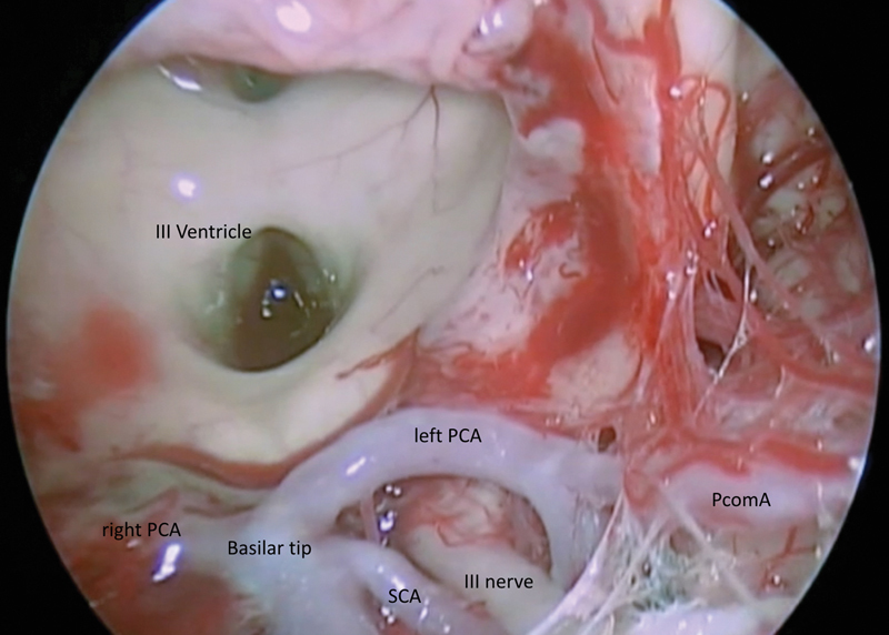
Exposure of the III ventricle and interpeduncular cistern after tumor resection. The basilar tip, PCA and SCA, III nerve, and PcomA are visible. PCA, posterior cerebral; PcomA, posterior communicating artery; SCA, superior cerebellar.

Exposure of the III ventricle and interpeduncular cistern after tumor resection. The basilar tip, PCA and SCA, III nerve, and PcomA are visible. PCA, posterior cerebral; PcomA, posterior communicating artery; SCA, superior cerebellar.