Figure 1.
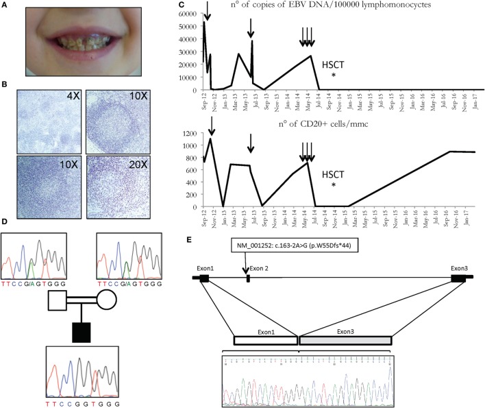
(A) Multiple caries in CD70 deficient patient. (B) Histologic analysis of patient’s lymph node showing follicular hyperplasia with paracortical expansion (top left), germinal centers with normal appearance (top right), follicular lysis (lower panels). (C) Copies of EBV DNA (above) and number of CD20 cells (below) detected in the patient. Arrows represent the infusions of rituximab. (D) Family pedigree with the c.163-2A>G variant of the CD70 gene, heterozygous in carriers, and homozygous in the patient. (E) CD70 genomic region and cDNA sequence of the first 3 exons in the patient is depicted, revealing the exon 2 skipping in the electropherogram at the bottom.

