
Fig 6.
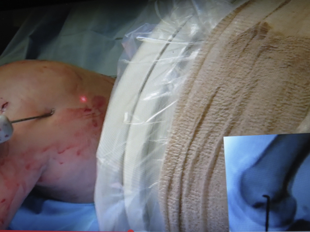
Right knee, exterior view of the distal medial femur. While the knee is flexed to avoid interference from the contralateral knee, the location of the medial epicodyle is identified fluoroscopically to assist in creation of an incision that allows access to the MCL's femoral attachment site. (MCL, medial collateral ligament.)