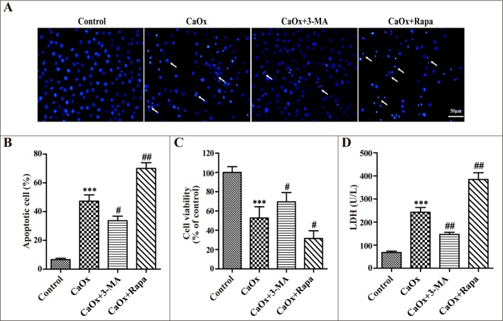
Figure 5. Effects of 3-methyladenine and rapamycin on CaOx crystal-induced HK-2 cell injury.
HK-2 cells were incubated with CaOx crystals (4 mM) for 24 h in the absence or presence of 3-methyladenine (3-MA, 5 mM) or rapamycin (Rapa, 10 μM). (A) DAPI staining was used to examine cell and nuclear morphology to analyze apoptosis. White arrows indicated apoptosis of HK-2 cells; scale bar: 50 μm. (B) Quantitative analysis of CaOx crystals induced apoptosis. (C) Cell viability was measured by CCK-8 assay. (D) The levels of LDH in culture supernatant were determined using the LDH assay. Data are presented as the mean ± SD from three experiments. ***P < 0.001 versus the control group, #P < 0.05, ##P < 0.01 versus the CaOx (4 mM) group.

