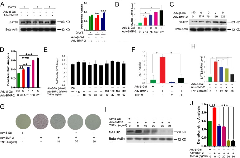
Figure 2. TNF-α inhibits osteoblastogenesis and SATB2 expression.
(A) The C2C12 cells were treated with Adv-BMP2 (150 pfu/cell) or Adv-β-Gal (150 pfu/cell) for one, three, and five days and SATB2 expression was examined by western blot and then underwent densitometric analysis. (B, C, D) The C2C12 cells were treated with Adv-β-Gal (150 pfu/cell) or various concentrations of Adv-BMP2 for three days. The SATB2 relative mRNA levels (B) and protein levels (C) were assessed and densitometric analysis of C (D) was graphed. (E) The C2C12 cells were treated with Adv-β-Gal (150 pfu/cell), Adv-BMP2 (150 pfu/cell), or 10 ng/mL to 60 ng/mL of TNF-α for 72 h, then followed by MTT assay. (F) The C2C12 cells were treated with Adv-β-Gal (150 pfu/cell), Adv-BMP2 (150 pfu/cell), or 60 ng/mL of TNF-α for five days. ALP activity were measured. (G) The C2C12 cells were treated with Adv-β-Gal (150 pfu/cell), Adv-BMP2 (150 pfu/cell), or 10 ng/mL to 60 ng/mL of TNF-α for five days and ALP staining was performed. (H_J) The C2C12 cells were treated with Adv-β-Gal (150 pfu/cell), Adv-BMP2 (150 pfu/cell), or 10 ng/mL to 60 ng/mL of TNF-α for 72 h. The SATB2 gene expressions were assessed by real-time PCR (H) and western blot (I) and densitometric analysis (J). The data are presented as mean ± S.D. (n = 3; *p < 0.05; **p < 0.01; ***p < 0.001), these western blot images were uncropped.

