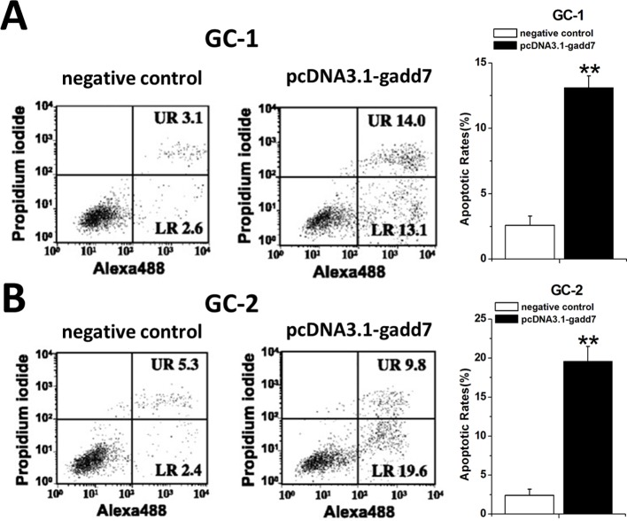
Figure 3. Involvement of gadd7 in cell apoptosis.
GC-1 and GC-2 cells were transfected with the plasmids in 6-well plates. Cell apoptosis was measured by the flow cytometry at 48 h post transfection. Representative images of flow cytometry analysis in GC-1 cells (A) and GC-2 cells (B) were shown. Cell apoptosis induction was observed in pcDNA3.1-gadd7 transfected GC-1 and GC-2 cells using flow cytometry analysis. Error bars, standard deviation. **P<0.01, compared with the negative control. The apoptosis differences were analyzed using independent samples t-test.

