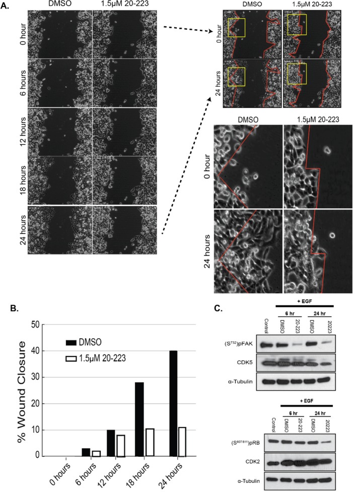
Figure 4. 20-223 disrupts migration of CRC cells.
(A) Wound gap images taken during the 24 hour incubation of HCT116 cells with DMSO or 1.5μM 20-223. 0 and 24 hour images were further evaluated by outlining the wound area (red lines) and zooming in on the wound boundaries (yellow box). (B) Quantification of % wound closure after treatment of HCT116 cells with DMSO of 1.5μM 20-223. (C) Western blot analyses at 6 and 24 hours after stimulation with EGF and treatment with either DMSO or 1.5μM 20-223.

