Figure 1.
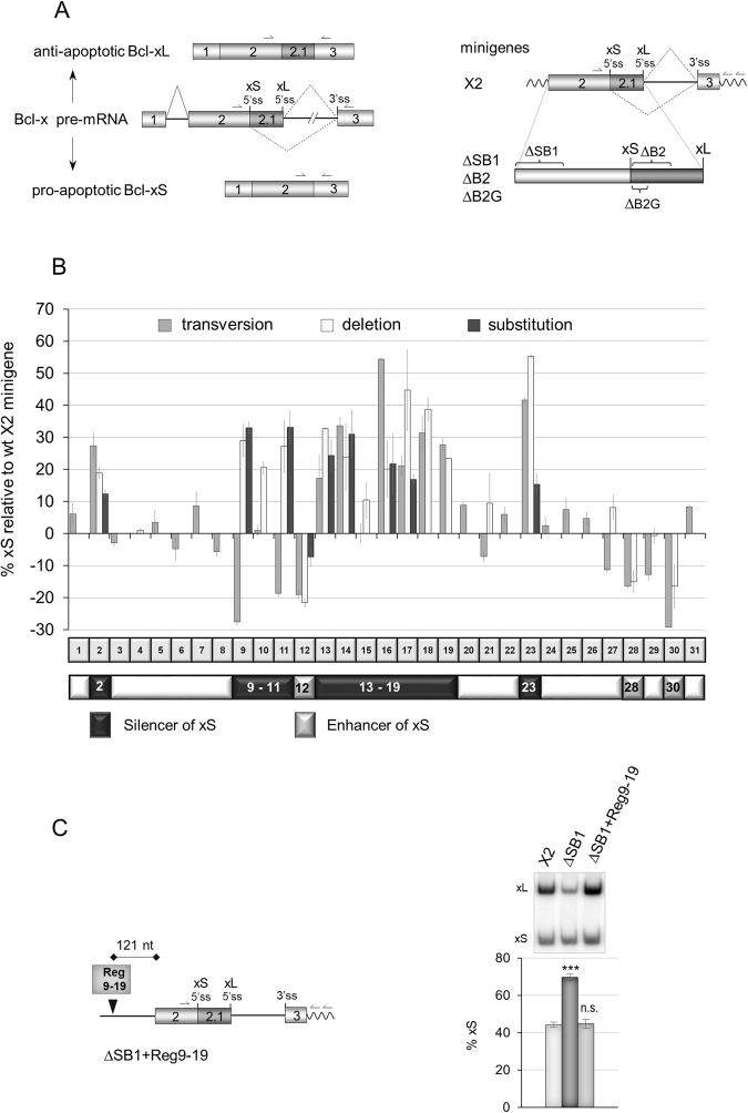
Mutagenesis of the SB1 element in Bcl-x exon 2. (A) Structure of the human Bcl-x pre-mRNA and its two splice variants anti-apoptotic Bcl-xL and the shorter pro-apoptotic isoform Bcl-xS. The portion included in minigene X2 and deleted in minigenes ΔSB1, ∆B2 and ∆B2G is shown on the right. Primer positions for RT-PCR assays from endogenous and minigene Bcl-x transcripts are shown. (B) The graph plots the percentage of Bcl-xS mRNA obtained by RT-PCR analysis for each mutation relative to the wild-type X2 minigene. The SB1 element was divided into 31 segments of 10 nt. One round of mutations changed A, G, C and T into T, C, G and A, respectively (transversions). A second set of mutations produced deletions of selected segments. Finally, specific segments were mutated by changing the wild-type sequence for GACTCAGTGT. Categorization or segments as displaying enhancer or silencer activity only considers the impact of deletions greater than 10 percentage points. (C) RT-PCR assays on total RNA extracted from ECR293 cells transfected with X2, ∆SB1 and ∆SB1 + Reg9-19 minigenes. The Reg9-19 region was inserted 279 nucleotides upstream of Bcl-xS 5′ss. The percentage of Bcl-xS is an average of triplicates. Error bars indicate SD. Asterisks represent significant P values (two-tailed Student’s t test) comparing the means between samples and their respective controls. *P < 0.05, **P < 0.01 and ***P < 0.001; n.s. = not significant.

