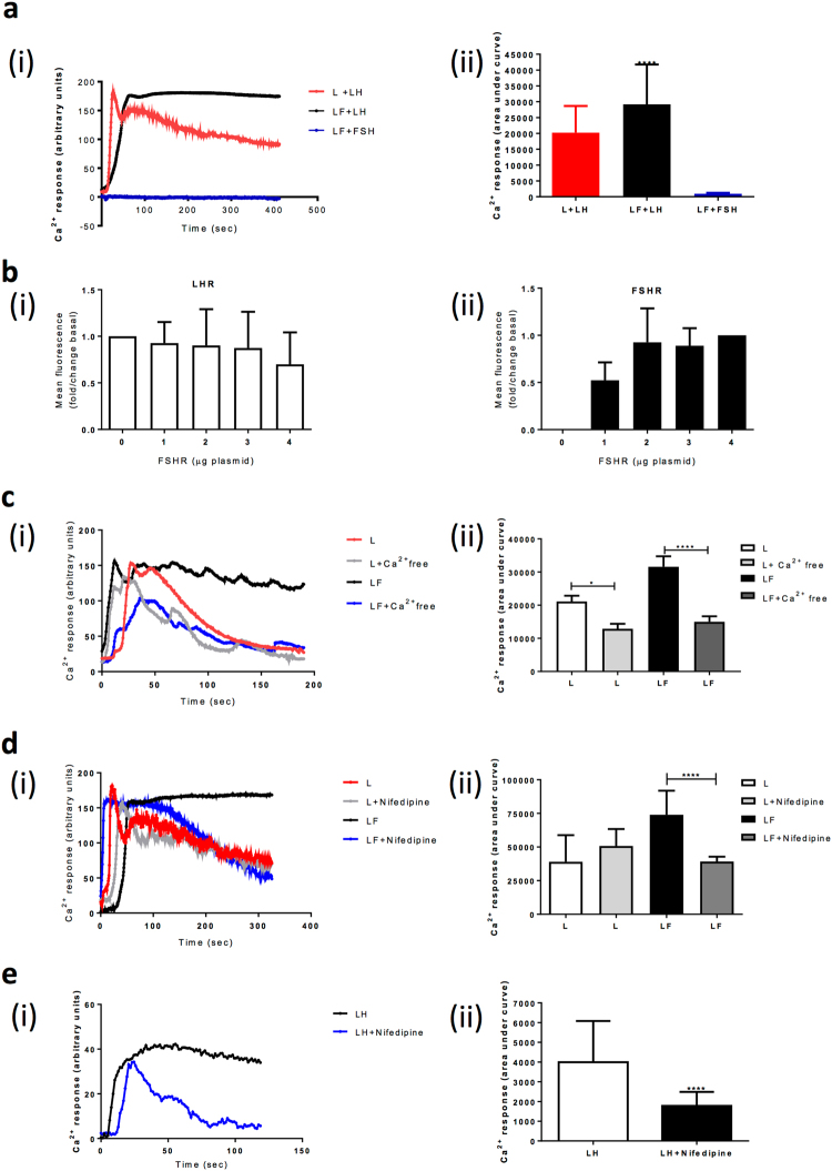
Figure 1.
LHR/FSHR crosstalk stimulates a sustained LH-dependent Ca2+ release that is dependent on extracellular Ca2+. (a) (i) Cells were treated with Ca2+ indicator dye Fluo4-AM and imaged live via confocal microscopy. Representative Ca2+ traces showing Ca2+ release in cells stably expressing LHR alone (L), or co-expressing LHR/FSHR (LF) in response to LH or FSH (100 nM). Representative fluorescent trace over time with baseline fluorescence subtracted. Ligand is added at t = 0. (ii) Quantitative analysis of data as described in a(i). Results are expressed as area under the curve from >20 cells per condition carried out in duplicate, n = 5. Data is presented as Mean ± SEM. (b) Measurement of cell surface expression via flow cytometry of (i) FLAG-tagged LHR and (ii) HA-tagged FSHR, in cells stably expressing FLAG-LHR and transiently transfected with increasing amounts of HA-FSHR plasmid DNA. Mean ± SEM, n = 5 (c) Assessment of the sustained LH-dependent Ca2+ response in cells co-expressing LHR/FSHR under Ca2+ free PBS conditions. Cells were incubated in Fluo4-AM indicator dye in Ca2+ free PBS prior to live confocal imaging and stimulation with LH (100 nM). (d) Measurement of LH-dependent Ca2+ responses with and without pre-treatment with nifedipine (10 μM, 30 min). (e) Assessment of LH-dependent Ca2+ responses in primary human granulosa-lutein cells co-expressing LHR/FSHR treated as in (d). Representative fluorescent traces over time with baseline fluorescence subtracted are shown in c(i)–e(i). For a(ii) and c(ii)–e(ii), results are expressed as area under the curve from n = 3–5 experiments, *p < 0.05, ****p < 0.0001.

