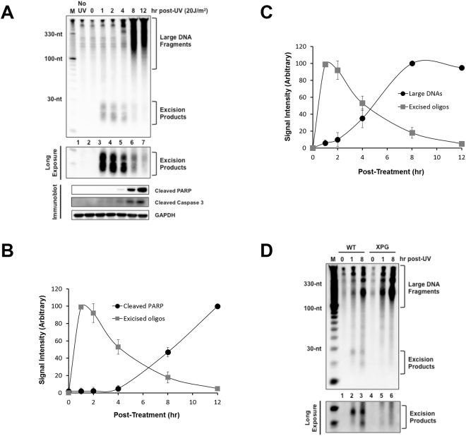
Figure 1.
Correlation between apoptotic signaling and the generation of large DNAs with decreased generation of excised oligonucleotide repair products. (A) Analysis of large DNA fragments and excised oligomers released from genomic DNA following UV irradiation. HeLa cells were exposed to 20 J/m2 of UVC and incubated for the indicated time points. After extraction of low molecular weight of DNAs from the cells, the DNA molecules were labeled with biotin at 3′-end using terminal deoxynucleotidyl transferase (TdT). The labeled DNA molecules were separated by gel electrophoresis, transferred to a nylon membrane and immobilized by UV cross-linking. The membrane was then incubated with HRP-conjugated streptavidin, and the labeled DNA molecules were detected with a chemiluminescence reagent (upper panel). A small portion of the UV-irradiated cells were lysed and analyzed by SDS-PAGE and immunoblotting with the indicated antibodies. (B) Quantification of the excised oligonucleotide repair products and cleaved PARP signals as shown in (A). The relative signal intensities were determined by setting the highest signal to 100. The results were quantified and are plotted as mean and standard deviations. (C) Quantitative analysis of the excised repair products and the large DNA molecules. (D) Wild-type (AA8) and XPG mutant (UV135) CHO cell lines were exposed to UV and processed as described above for the detection of soluble DNAs.

