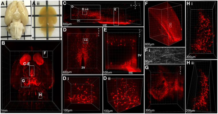
Figure 1.
Whole-mount immunolabelling and optical tissue clearing in the rat brain. (A) The intact rat brain before (i) after (ii) immunolabelling for tyrosine hydroxylase (TH) and tissue clearing. (B) 3D rendering of TH-immunoreactivity (ir) in the rat brain imaged in the horizontal plane and viewed from the ventral surface (z depth = 4 mm). (C) Hypothalamic TH-ir neurons viewed in the sagittal plane from −2.6 mm to −4 mm Bregma. Insets (D,E): (D) TH-ir neurons in periventricular nuclei viewed from the dorsal surface with high magnification renderings of neurons in the horizontal plane (D i) and rotated to the sagittal (D ii) plane. (E) TH-ir neurons within a portion of the dorsomedial and arcuate hypothalamic nuclei projected into the coronal plane. (F) 3D rendering and a high resolution projected image (F i) of TH-ir fibers in the cortex. (G) TH-positive neurons within the substantia nigra and ventral tegmental area of the midbrain. (H) TH-positive neurons of the A5 group located in the ventrolateral pons viewed in the horizontal plane (i) and rotated into the sagittal plane (ii). R = rostral, C = caudal, D = dorsal, V = ventral, oc = optic chiasm, ME = median eminence, 3 V = third ventricle.

