Fig. 1.

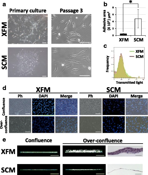
Morphological appearance and morphometric analysis of DPSCs under xenogeneic serum-free or FBS-containing culture conditions. a Phase-contrast images of DPSCs cultured in XFM or SCM in primary culture. Scale bars, 200 μm. Passage 3: scale bars, 100 μm. b Cell morphometric evaluation of adhesive areas of XFM and SCM cells at passage 3. *P < 0.01. c Flow-cytometric analysis of single-cell size in XFM and SCM cellular suspensions. d Phase-contrast (Ph) and fluorescence-microscope images of DAPI-labeled XFM and SCM cells at confluence on day 10 or overconfluence on day 14 post seeding. Scale bars, 100 μm. e 3D fluorescence imaging and HE staining of confluent and overconfluent cultures of Phalloidin-labeled XFM and SCM cells. Scale bars, 100 μm. SCM xenogeneic serum-containing culture medium, XFM xenogeneic serum-free culture medium, DAPI 4′,6-diamidino-2-phenylindole
